My Research Project Was Scrapped Due to Lack of Tissue
 I always knew I wanted to be a scientist. This meant that when I was younger, I loved watching movies and shows featuring some scientific aspect. Science is usually shown as someone having an idea, immediately rushing to the the lab, and finding out a result that day. Science in real life, however, requires months of planning to see if the idea is good, more planning on how to test it, and way too long of a publishing process.
Science is slow and arduous, it can go wrong at any step of the way. The first steps of science are pretty simple, observe something you’re interested in and ask a question about it. Next, you need funding and supplies. Funding is often in short supply in pediatric oncology, but there are times you’re lucky and find enough.
Materials are next, normally these materials are already in the lab or ordered. Except for tissue. Tissue comes in all types and from all types of organisms but it is a finite resource. And absolutely necessary. If an experiment calls for tissue, no matter how small the amount, there is no substitute for it.
This can create a huge obstacle for research advances. Science is slow enough to begin with, having the process held up any more is not ok. Especially in oncological research, where translating your findings as quickly as possible into treatments can have a direct impact on lives. The more rare the disease, the less tissue there is available. Rare doesn’t mean less devastating or less deserving of research. But it can mean impeded research.
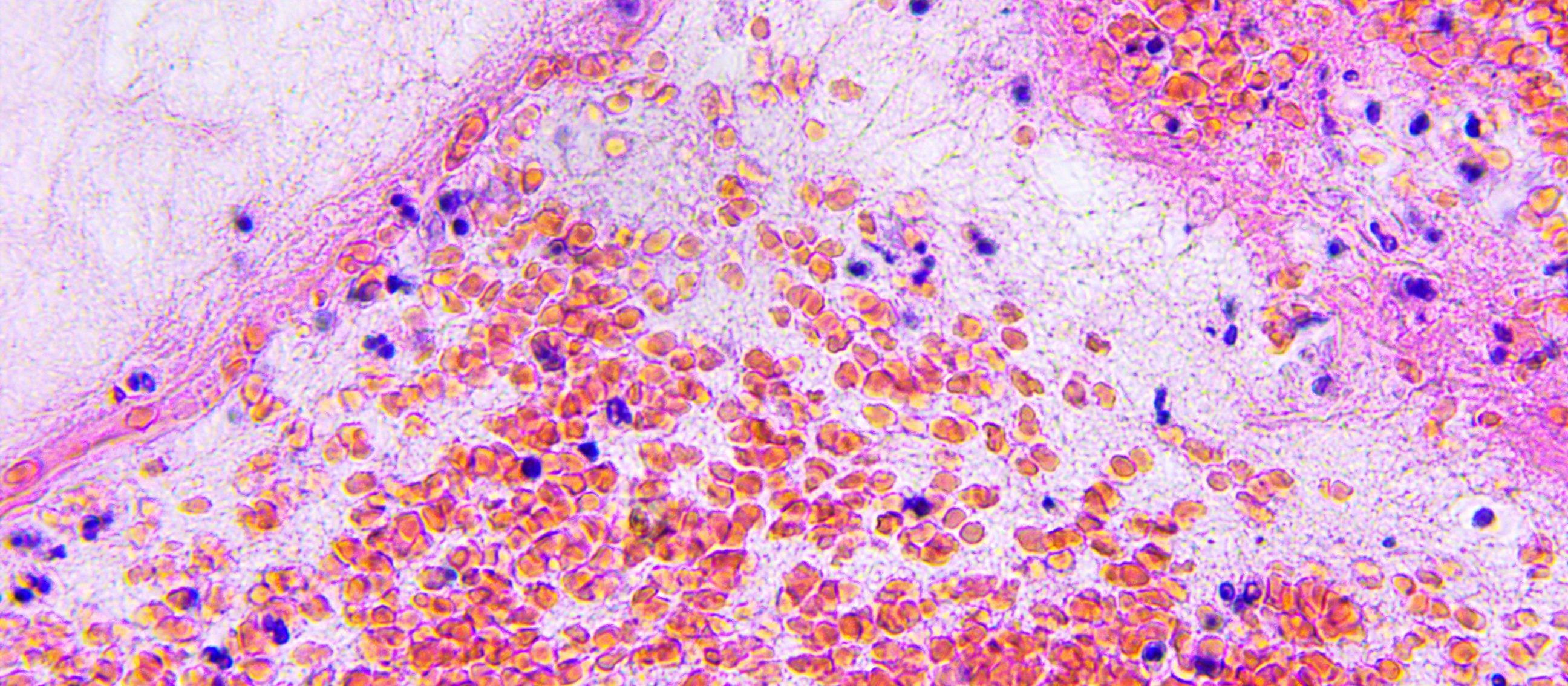
 I ran into this problem at my first ever research position. My first independent project was going to be on a rare pediatric brain tumor called a Craniopharyngioma. I thought I was all set to go, I had previous data to work with, funding, and a lab full of supplies. What I was missing was tissue. I first tried ordering the tissue samples of frozen tumors that you “wake up” in the lab. There are several websites to use for this, none of them had what I needed.
I then decided to see what others had done in my situation. I read dozens and dozens of papers on this tumor, trying to find recent ones that used tissue rather than retroactive data. Majority of these papers were case studies, they had gotten their tissue directly from a singular patient and that was it. The few articles I found where the authors were in a similar situation, I emailed the authors. I emailed scientists from all over the states, Germany, China, Brazil, and no one had tissue that was available and stored in the correct way to send it to me.
This went on for weeks but ultimately my project was scrapped due to lack of tissue.
This is why GFAC is so important. Not even close to enough researchers are looking at pediatric brain tumors, there have only been 3 drugs in over 40 years developed for pediatric patients. There is less focus because they are considered rare.
But again, rare does not mean less deserving. With GFAC changing the culture surrounding post-mortem tissue donation, it means fewer projects scrapped like mine was, better access to tissue, and science turning more quickly into treatments. More lives are saved this way.
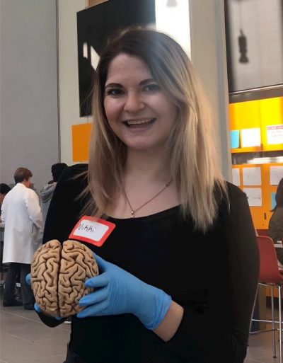
Nikki Lyons is a neuroscience and molecular biology student at Columbia University in NYC.She works as a pediatric cancer researcher and advocate and is planning on becoming a surgical neuro-oncologist. Nikki has been an intern with the Children’s Brain Tumor Tissue Consortium in Philadelphia and a research assistant at Ann and Robert Lurie Children’s Hospital in Chicago. She currently interns with Swifty Foundation and Gift from a Child.
Recent Publications from Centers of Excellence
Harmonization of Post-mortem Donations for Pediatric Brain Tumors and Molecular Characterization of Difuse Midline Gliomas
Children diagnosed with brain tumors have the lowest overall survival of all pediatric cancers. To address the paucity of tissue for biological studies, we have established a comprehensive protocol for the coordination and processing of donated specimens at postmortem. Since 2010, 60 postmortem pediatric brain tumor donations from 26 institutions were coordinated and collected. Patient derived xenograft models and cell cultures were successfully created (76% and 44% of attempts respectively), irrespective of postmortem processing time. Histological analysis of mid-sagittal whole brain sections revealed evidence of treatment response, immune cell infiltration and the migratory path of infiltrating H3K27M DMG cells into other midline structures and cerebral lobes. Sequencing of primary and disseminated tumors confirmed the presence of oncogenic driver mutations and their obligate partners. Our findings highlight the importance of postmortem tissue donations as an invaluable resource to accelerate research, potentially leading to improved outcomes for children with aggressive brain tumors. Read Full Publication
 Dr. Monje-Deisseroth and her team at Stanford University recently published a paper detailing how gliomas are able to “hijack” the brain's communication system.
Published in Nature: High-grade gliomas are lethal brain cancers whose progression is robustly regulated by neuronal activity. Activity-regulated release of growth factors promotes glioma growth, but this alone is insufficient to explain the effect that neuronal activity exerts on glioma progression. Here we show that neuron and glioma interactions include electrochemical communication through bona fide AMPA receptor-dependent neuron–glioma synapses. Read More
Congratulations to two of our Center of Excellence teams lead by Dr Javad Nazarian and Dr. Michelle Monje on their recent groundbreaking research publication. Due in part to increased access to post-mortem tissue the teams were able to study a larger sample of DIPG tumors. Diffuse intrinsic pontine glioma is a lethal pediatric brain cancer characterized by H3K27M histone mutation. Nagaraja et al. characterize a large cohort of rare primary tumors and normal pontine tissue to reveal active regulatory element heterogeneity dependent upon the histone variant and cell context in which the mutation occurs. Read More
Research Breakthroughs Resulting from Autopsy Tissue
Donovan_et_al_Locoregional_delivery_CAR-T_cells_Nat_Med2020_opt
Potent antitumor efficacy of anti-GD2 CAR T cells in H3-K27M+ diffuse midline gliomas_2018
Non-inflammatory tumor microenvironment of diffuse intrinsic pontine glioma_2018
Intertumoral Heterogeneity within Medulloblastoma Subgroups_Sick Kids Publication_June 2017
A Protocol for Rapid Post-mortem Cell Culture of Diffuse Intrinsic Pontine Glioma (DIPG)_March 2017
Neural Precursor-Derived Pleiotrophin Mediates Subventricular Zone Invasion by Glioma_Aug 2017
Divergent clonal selection dominates medulloblastoma at recurrence
Models Pave the Way for Improved Outcomes in Medulloblastoma_August 2016
Functionally defined therapeutic targets in diffuse intrinsic pontine glioma_June 2015
Neuronal Activity Promotes Glioma Growth through Neuroligin-3 Secretion_May 2015 Cell
Stanford Research Paper: Electrical and synaptic integration of glioma into neural circuits
Rapid autopsy of a patient with recurrent anaplastic ependymoma.
Next-Generation Rapid Autopsies Enable Tumor Evolution Tracking and Generation of Preclinical Models
Why Autopsy Tissue is Needed to Empower Research
Pediatric Brain Cancer Tissue Donation article_2016
Overcoming Autopsy Barriers in Pediatric Cancer Research
Study on Collection of autopsy tissue in DIPG patients_Oct 2010
Next-Generation Rapid Autopsies Enable Tumor Evolution Tracking_January 2018
The clinical, research, and social value of autopsy after any cancer death
Gift from a Child
Is a Swifty Foundation Program
Swifty is a recipient of the GuideStar Gold Seal of Transparency