Research
Empowering Brain Cancer Research Through Tissue Donation
Pediatric brain cancer research is not proceeding as fast as it could and survival rates have flatlined because tissue for research is scarce.
The Power of Tissue Donation

Autopsy Tissue
=

Empowered Research
=

Medical Advances
The precious gift of your child’s autopsy tissue will empower research so breakthroughs can be made and a cure can be found for this devastating disease. Here you will find a listing of some pediatric cancer publications that 1) explain the need for autopsy tissue and 2) highlight some of the research breakthroughs that have been made possible because of autopsy tissue. *Some articles have been renamed for those of us that are not STEM professionals.
Recent Publications from Centers of Excellence
The Landscape of Tumor Cell States and Spatial Organization in H3-K27M Mutant Diffuse Midline Glioma Across Age and Location
Donated tissue is an informative tool in medical research.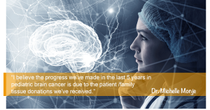
Among the many methods scientists use to understand human diseases, donated patient tissue is a uniquely precious tool. A recent study published this month in Nature Genetics investigated the heterogeneity of cell types and gene expression in DIPG/DMG patient tissue possessing the characteristic H3K27M histone mutation. Dr. Mariella Filbin’s team, in collaboration with Dr. Mats Nilsson in Stockholm and Dr. Michelle Monje at Stanford (a GFAC Center of Excellence), utilized patient tissue donated across several years to analyze the cellular composition of these aggressive pediatric tumors. Previous research has suggested these tumors arise not from neurons, but from cells resembling oligodendrocyte precursor cells (OPCs). OPCs have many important functions in the brain throughout the lifespan. This study addresses ongoing questions in DMG research including: the gene expression profiles of these cancer cells, changes in expression between different brain region and patients, and the cellular arrangement of the tumor.
Postmortem Tissue Donation: Giving Families the Ability to Choose
Patti and Al Gustafson along with members of the GFAC recently published a consensus paper from the family perspective
Postmortem tissue donations can represent a driving force toward progress in brain tumor research, particularly malignant and recurrent tumors where current survival is dismal. In addition to the scientific value, there are benefits to postmortem tissue donation from the parental perspective. However, several real and perceived barriers exist, which have limited the rate of autopsy including discrepancies between clinician, patient, and parental views. To identify and address these barriers, a consensus conference of advocates of children who died of brain cancer was held in December 2018 at the Children’s Hospital of Philadelphia. Major barriers to obtaining autopsy were identified with an emerging theme that a cultural shift is urgently required where families and patients with terminal brain cancers are uniformly provided with the option of postmortem donation.
Summarizing the opinions collected from more than 120 parents whose children died of brain cancer and leveraging the experience of the multi-institutional postmortem CNS tumor collection program, Gift from a Child (GFAC), we seek to inform medical professionals of the need to approach families about this sensitive topic and to demonstrate how barriers to donation can be overcome. A central theme among parent advocates is that families and patients ultimately do want to be asked and provided the opportunity. Specifically, there was broad consensus that processes need to be embedded that require clinicians to broach the topic and ask all families, and a failure to ask robs them of an important opportunity
Love Never Dies
Through Gift From a Child, parents can turn loss and grief into action.
A recent collaborative effort by the entire Gift from a Child team was published in the National Funeral Directors Association summer newsletter. The project, lead by Melissa Williams, GFAC Tissue Navigator was part of our outreach efforts into adjacent organizations to educate and collaborate.
Medical journal articles report that many bereaved parents who have lost children to brain cancers are interested in supporting medical research through postmortem tissue donations. Their greatest hope is that families in the future will be spared the same loss because of scientific advancements enabled through these research donations. The overall program is meant to be gentle on families that make the decision to donate. They are provided with information about how the program
works and a general timeline of the process, and will sign a consent for the tissue donation (research autopsy).
Donation can take place wherever the death occurs – hospital, home or hospice – and the family is given all the time they want and need with their child at the time of death. Read Full Publication
Harmonization of Post-mortem Donations for Pediatric Brain Tumors and Molecular Characterization of Difuse Midline Gliomas
![]() An important study from our team at Children’s National. Children diagnosed with brain tumors have the lowest overall survival of all pediatric cancers. To address the paucity of tissue for biological studies, we have established a comprehensive protocol for the coordination and processing of donated specimens at postmortem. Since 2010, 60 postmortem pediatric brain tumor donations from 26 institutions were coordinated and collected. Patient derived xenograft models and cell cultures were successfully created (76% and 44% of attempts respectively), irrespective of postmortem processing time. Histological analysis of mid-sagittal whole brain sections revealed evidence of treatment response, immune cell infiltration and the migratory path of infiltrating H3K27M DMG cells into other midline structures and cerebral lobes. Sequencing of primary and disseminated tumors confirmed the presence of oncogenic driver mutations and their obligate partners. Our findings highlight the importance of postmortem tissue donations as an invaluable resource to accelerate research, potentially leading to improved outcomes for children with aggressive brain tumors. Read Full Publication
An important study from our team at Children’s National. Children diagnosed with brain tumors have the lowest overall survival of all pediatric cancers. To address the paucity of tissue for biological studies, we have established a comprehensive protocol for the coordination and processing of donated specimens at postmortem. Since 2010, 60 postmortem pediatric brain tumor donations from 26 institutions were coordinated and collected. Patient derived xenograft models and cell cultures were successfully created (76% and 44% of attempts respectively), irrespective of postmortem processing time. Histological analysis of mid-sagittal whole brain sections revealed evidence of treatment response, immune cell infiltration and the migratory path of infiltrating H3K27M DMG cells into other midline structures and cerebral lobes. Sequencing of primary and disseminated tumors confirmed the presence of oncogenic driver mutations and their obligate partners. Our findings highlight the importance of postmortem tissue donations as an invaluable resource to accelerate research, potentially leading to improved outcomes for children with aggressive brain tumors. Read Full Publication
 Dr. Monje-Deisseroth and her team at Stanford University recently published a paper detailing how gliomas are able to “hijack” the brain’s communication system.
Published in Nature: High-grade gliomas are lethal brain cancers whose progression is robustly regulated by neuronal activity. Activity-regulated release of growth factors promotes glioma growth, but this alone is insufficient to explain the effect that neuronal activity exerts on glioma progression. Here we show that neuron and glioma interactions include electrochemical communication through bona fide AMPA receptor-dependent neuron–glioma synapses. Read More
Congratulations to two of our Center of Excellence teams lead by Dr Javad Nazarian and Dr. Michelle Monje on their recent groundbreaking research publication. Due in part to increased access to post-mortem tissue the teams were able to study a larger sample of DIPG tumors. Diffuse intrinsic pontine glioma is a lethal pediatric brain cancer characterized by H3K27M histone mutation. Nagaraja et al. characterize a large cohort of rare primary tumors and normal pontine tissue to reveal active regulatory element heterogeneity dependent upon the histone variant and cell context in which the mutation occurs. Read More
Research Breakthroughs Resulting from Autopsy Tissue
*Some titles have been changed for general public understanding
Study of CAR T cells for the treatment of metastatic medulloblastoma and ependymoma
Possible immunotherapy strategy for use in diffuse midline gliomas
Non-inflammatory tumor microenvironment of diffuse intrinsic pontine glioma_2018
Intertumoral Heterogeneity within Medulloblastoma Subgroups_Sick Kids Publication_June 2017
A Protocol for Rapid Post-mortem Cell Culture of Diffuse Intrinsic Pontine Glioma (DIPG)_March 2017
Potential therapeutic targets to limit glioma invasion.
Divergent clonal selection dominates medulloblastoma at recurrence
Models Pave the Way for Improved Outcomes in Medulloblastoma_August 2016
Functionally defined therapeutic targets in diffuse intrinsic pontine glioma_June 2015
The Important role of active neurons in promoting glioma growth
Rapid autopsy of a patient with recurrent anaplastic ependymoma.
Next-Generation Rapid Autopsies Enable Tumor Evolution Tracking and Generation of Preclinical Models
Why Autopsy Tissue is Needed to Empower Research
Pediatric Brain Cancer Tissue Donation article_2016
Overcoming Autopsy Barriers in Pediatric Cancer Research
Tissue Banking in Age of Precision Medicine
Study on Collection of autopsy tissue in DIPG patients_Oct 2010
Next-Generation Rapid Autopsies Enable Tumor Evolution Tracking_January 2018
The clinical, research, and social value of autopsy after any cancer death
Gift from a Child
Is a Swifty Foundation Program
Swifty is a recipient of the GuideStar Gold Seal of Transparency